-
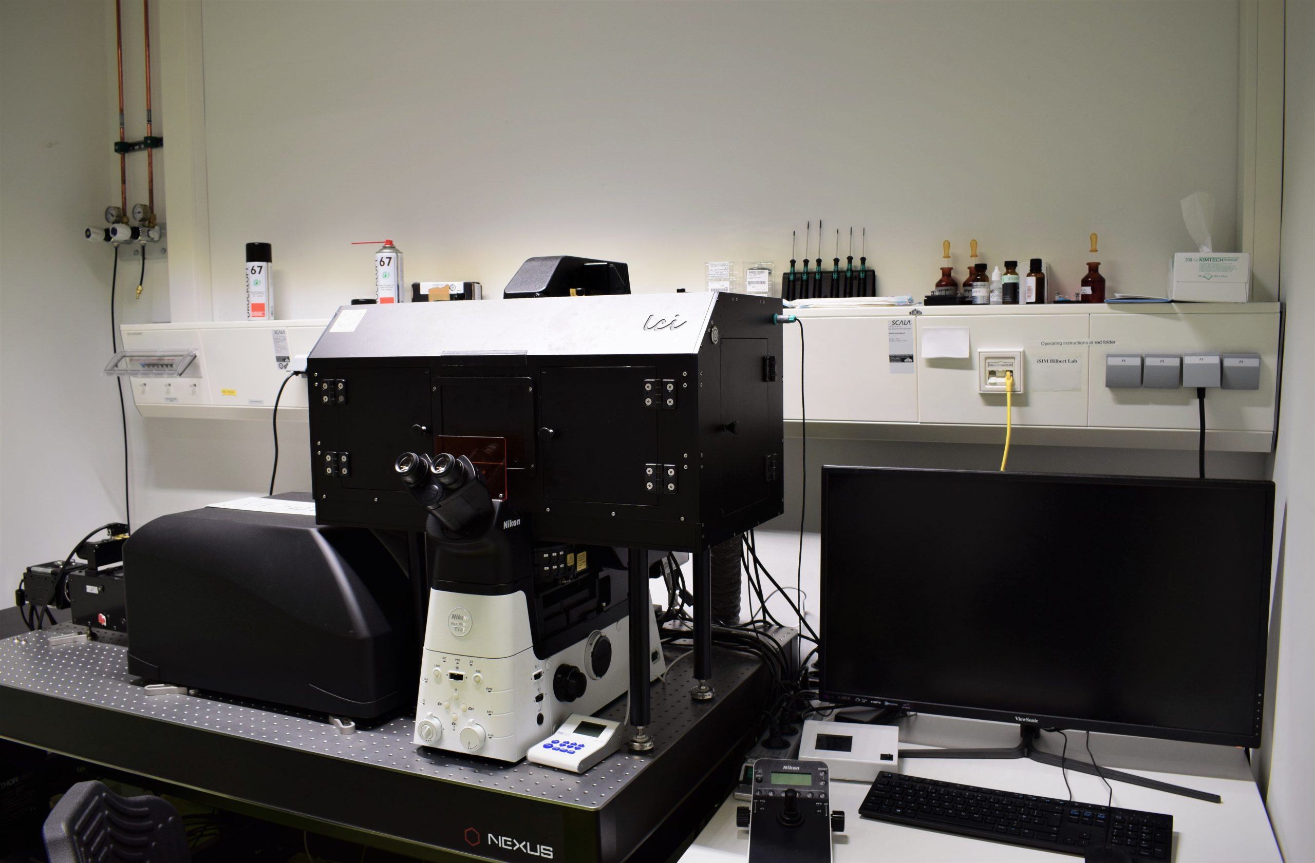
Instant-SIM microscope

-
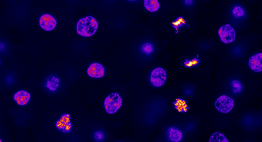
Chromatin distribution in nuclei of a pluripotent zebrafish embryo
The image shows the DNA distribution in different nuclei contained within a pluripotent zebrafish embryo. The image data were obtained with a Stimulated Emission Depletion (STED) super-resolution microscope at the Karlsruhe Center for Optics and Photonics by Lennart Hilbert. The image was used as the June 2021 issue cover for PLoS Computational Biology
-
Stages of Zebrafish Development
Illustrations of the stages “oblong”, “sphere”, “dome”, “30% epiboly” and “50% epiboly” of the embryonic development in zebrafish. At the reference temperature of 28.5°C, the developmental stages occur at the indicated time points, measured in hours post fertilization (hpf). Illustration by Xenia Tschurikow.
-
Visualisation of Nuclear Actin Bundles after Cell Division
The figure shows actin bundle formation after mitosis. Actin was visualized using recombinantly expressed actin chromobody with green fluorescent protein as a fluorescence marker and a nuclear localization sequence (NLS) to restrict detection to within the cell nucleus (nAC-GFP, generous gift from Grosse Lab, Uni Freiburg). Time-lapse images are single confocal sections chosen from z-stacks […]

